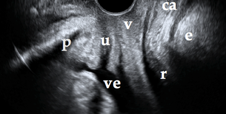
The two-dimensional ultrasound gives us a good anatomical image of the midsagittal plane. In this plane we can see the pubic symphysis, urethra, bladder, vagina, anal canal, rectum and the lowermost part of the levator ani muscle (Figure 1).
Figure 1. The two-dimensional cross section must include: the pubic symphysis (p), the urethra (u), the bladder (ve), the vagina (v), the anal canal (ac), the rectum (r) and the lowermost part of the levator ani muscle (l).
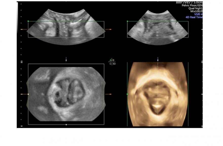
The three-dimensional ultrasound adds a third dimension that not seen in the two-dimensional ultrasound: the axial plane, which clearly depicts the urogenital hiatus, formed by the levator ani muscle. Until now, this plane could only be seen using MRI scans. This is the three-dimensional ultrasound's great contribution to the study of the pelvic floor (Figure 2).
Figure 2. The sagittal plane is shown at the top left; the transverse plane is shown on the right, the axial plane is shown on the left and the volumetric reconstruction of the urogenital hiatus can be seen on the right
Since it navigates through volume, the three-dimensional ultrasound offers us the possibility of looking at the same area from different angles. Using the sagittal, transverse and axial planes, we are offered a volumetric reconstruction of the muscles that form the pelvic floor. The 4-D ultrasound allows for dynamic imaging of volumes and can therefore capture images when the patient is performing the Valsalva manoeuvre or contracting the pelvic floor musculature; we can therefore navigate through a sequence of volumes until we find the volume that corresponds with maximum Valsalva or maximum contraction.